-
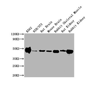
Western Blot
Positive WB detected in: K562 whole cell lysate, NIH/3T3 whole cell lysate, Rat Brain tissue, Mouse Brain tissue, Rabbit Skeletal Muscle tissue, Rat Kidney tissue, Rabbit Kidney tissue
All lanes ENO1 antibody at 1:10000
Secondary
Goat polyclonal to mouse IgG at 1/10000 dilution
Predicted band size: 47 KDa
Observed band size: 47 KDa
Exposure time: 1min
-
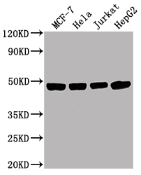
Western Blot
Positive WB detected in: MCF-7 whole cell lysate, Hela whole cell lysate, Jurkat whole cell lysate, HepG2 whole cell lysate
All lanes ENO1 antibody at 1:10000
Secondary
Goat polyclonal to mouse IgG at 1/10000 dilution
Predicted band size: 47 KDa
Observed band size: 47 KDa
Exposure time: 10s
-
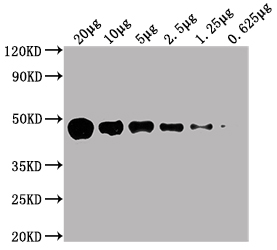
Western Blot
Positive WB detected in: HepG2 whole cell lysate at 20µg, 10µg, 5µg, 2.5µg, 1.25µg, 0.625µg
All lanes: ENO1 antibody at 1:5000
Secondary
Goat polyclonal to Mouse IgG at 1/10000 dilution
Predicted band size: 47 kDa
Observed band size: 47 KDa
Exposure time: 10s
-
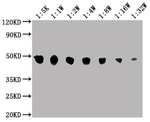
Western Blot
Positive WB detected in: MCF-7 whole cell lysate
All lanes: ENO1 antibody at 1:5000, 1:10000, 1:20000, 1:40000, 1:80000, 1:160000, 1:320000
Secondary
Goat polyclonal to Mouse IgG at 1/10000 dilution
Predicted band size: 47 kDa
Observed band size: 47 KDa
Exposure time: 10s
-
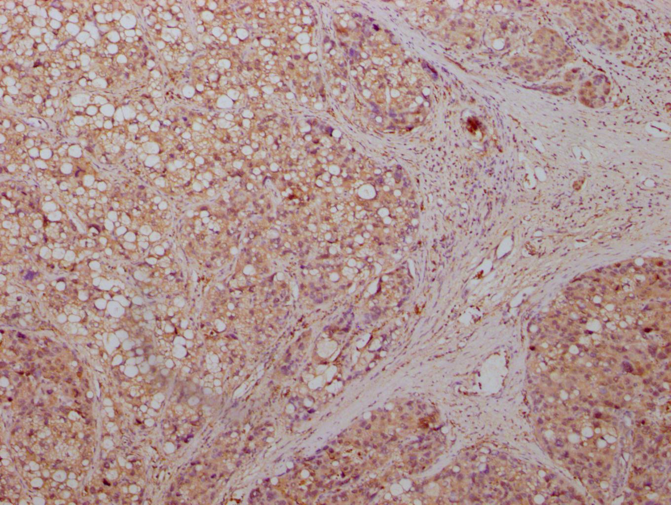
IHC image of CSB-MA007670A0m diluted at 1:500 and staining in paraffin-embedded human liver cancer tissue performed on a Leica BondTM system. After dewaxing and hydration, antigen retrieval was mediated by high pressure in a citrate buffer (pH 6.0). Section was blocked with 10% normal goat serum 30min at RT. Then primary antibody (1% BSA) was incubated at 4°C overnight. The primary is detected by a biotinylated secondary antibody and visualized using an HRP conjugated SP system.
-
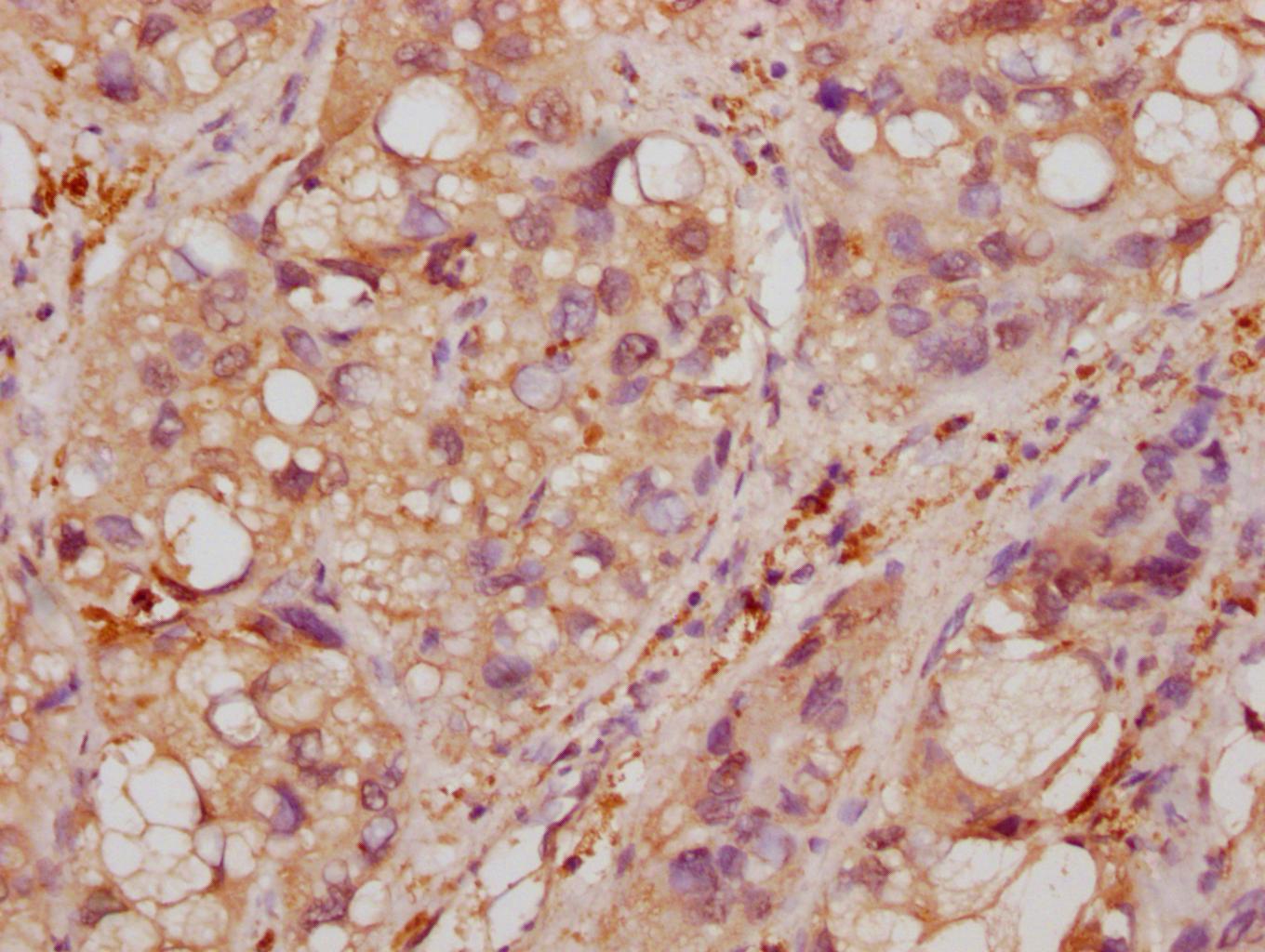
IHC image of CSB-MA007670A0m diluted at 1:500 and staining in paraffin-embedded human liver cancer tissue performed on a Leica BondTM system. After dewaxing and hydration, antigen retrieval was mediated by high pressure in a citrate buffer (pH 6.0). Section was blocked with 10% normal goat serum 30min at RT. Then primary antibody (1% BSA) was incubated at 4°C overnight. The primary is detected by a biotinylated secondary antibody and visualized using an HRP conjugated SP system.
-
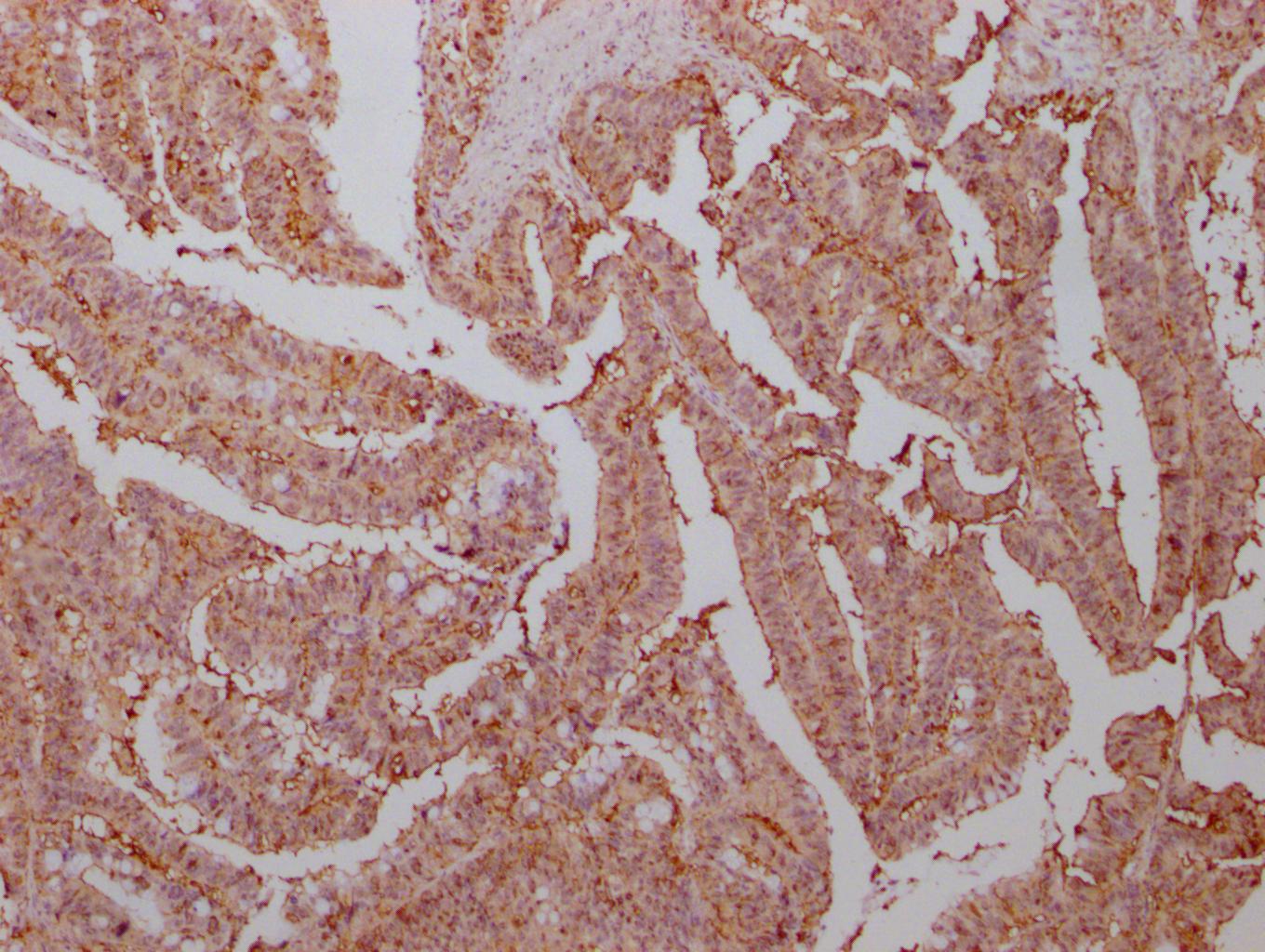
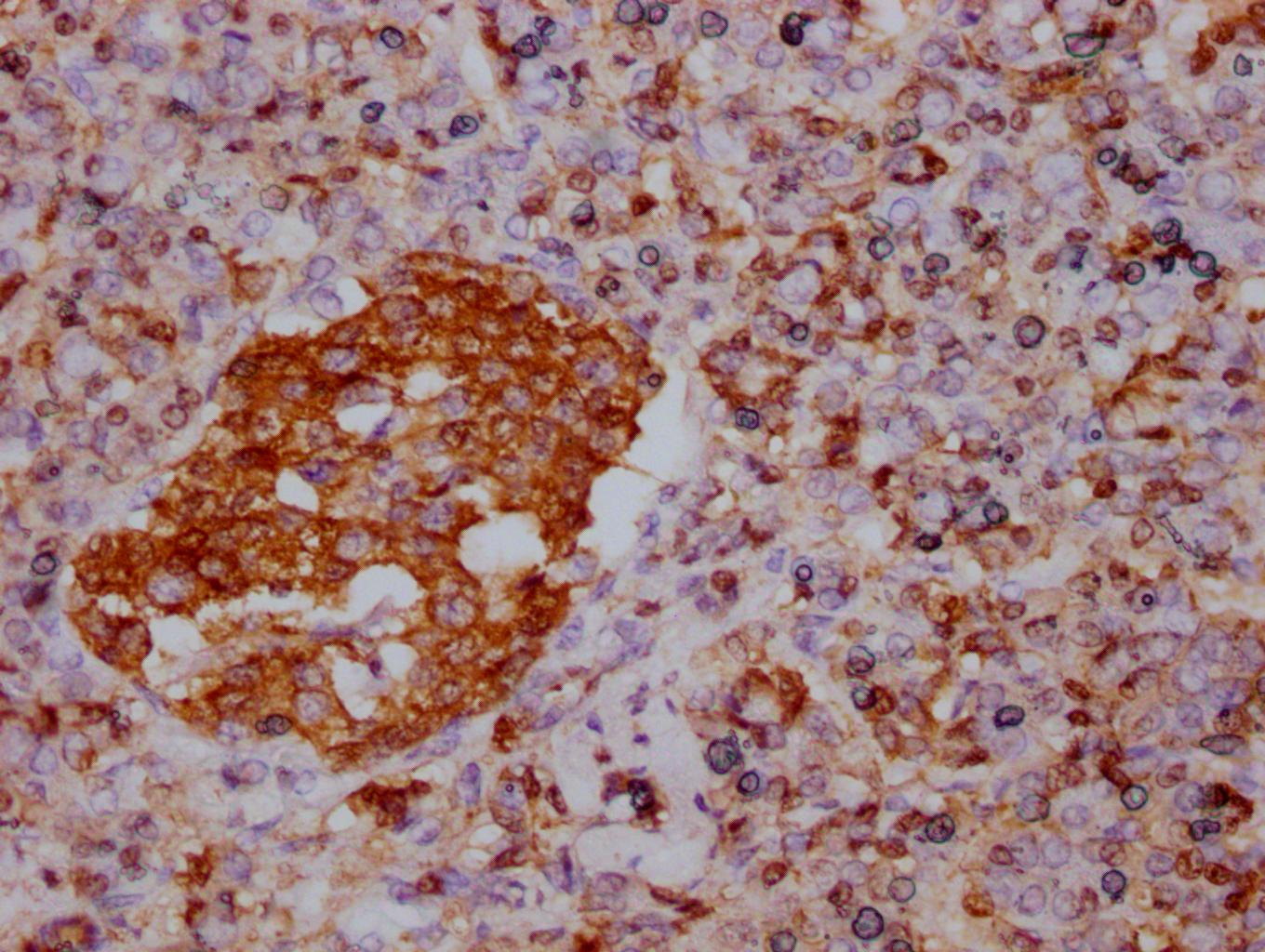
IHC image of CSB-MA007670A0m diluted at 1:500 and staining in paraffin-embedded human colon cancer tissue performed on a Leica BondTM system. After dewaxing and hydration, antigen retrieval was mediated by high pressure in a citrate buffer (pH 6.0). Section was blocked with 10% normal goat serum 30min at RT. Then primary antibody (1% BSA) was incubated at 4°C overnight. The primary is detected by a biotinylated secondary antibody and visualized using an HRP conjugated SP system.
-
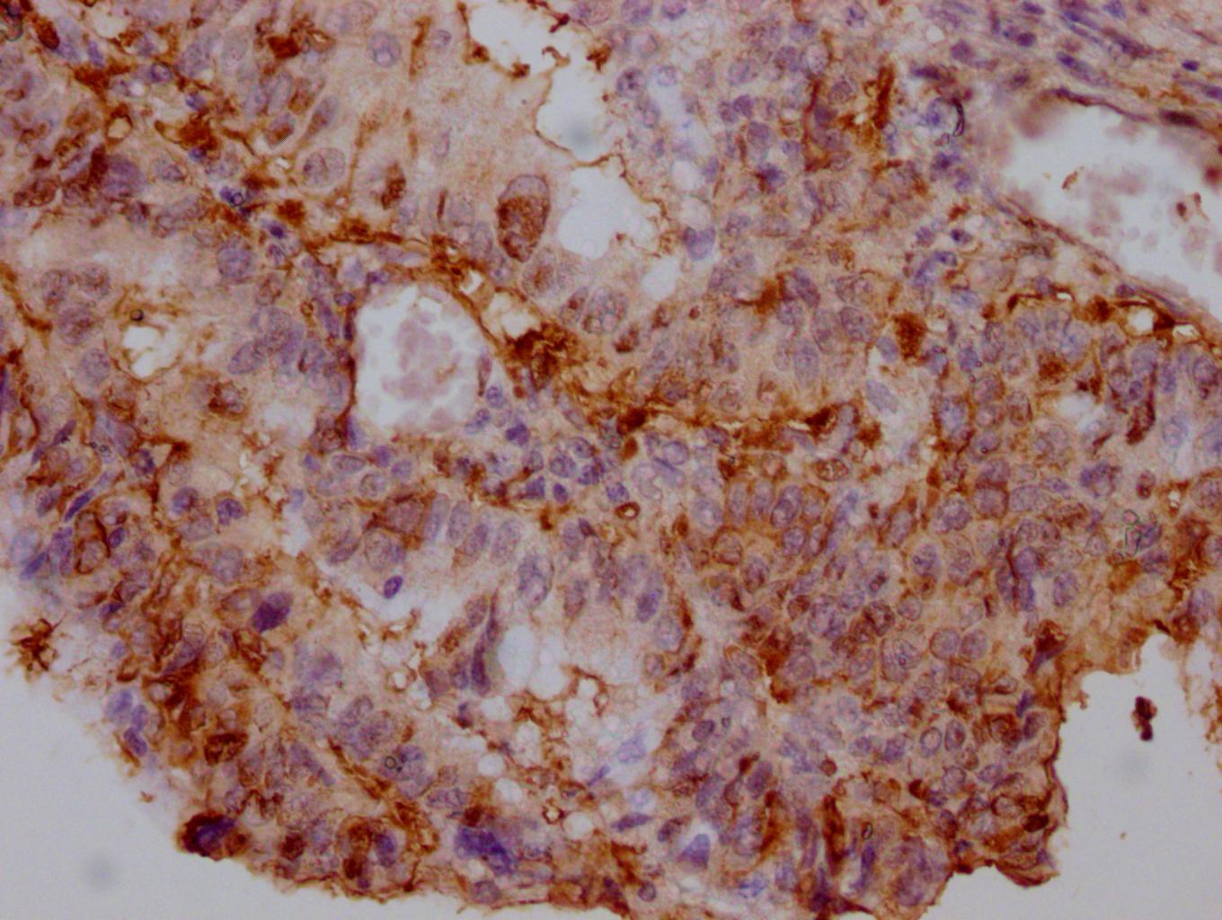
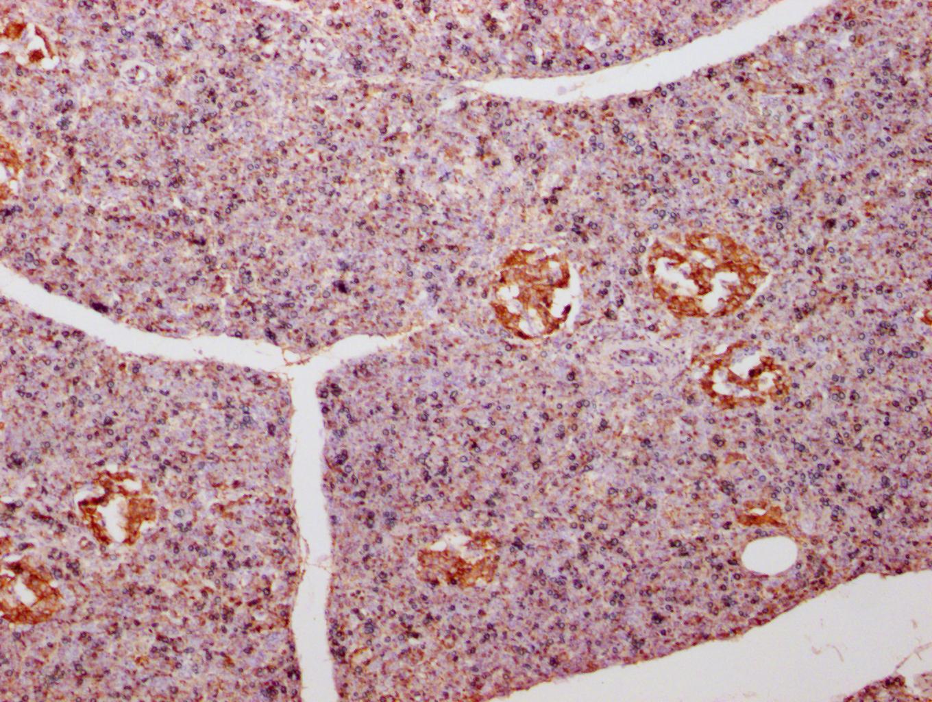
IHC image of CSB-MA007670A0m diluted at 1:500 and staining in paraffin-embedded human pancreas tissue performed on a Leica BondTM system. After dewaxing and hydration, antigen retrieval was mediated by high pressure in a citrate buffer (pH 6.0). Section was blocked with 10% normal goat serum 30min at RT. Then primary antibody (1% BSA) was incubated at 4°C overnight. The primary is detected by a biotinylated secondary antibody and visualized using an HRP conjugated SP system.
-
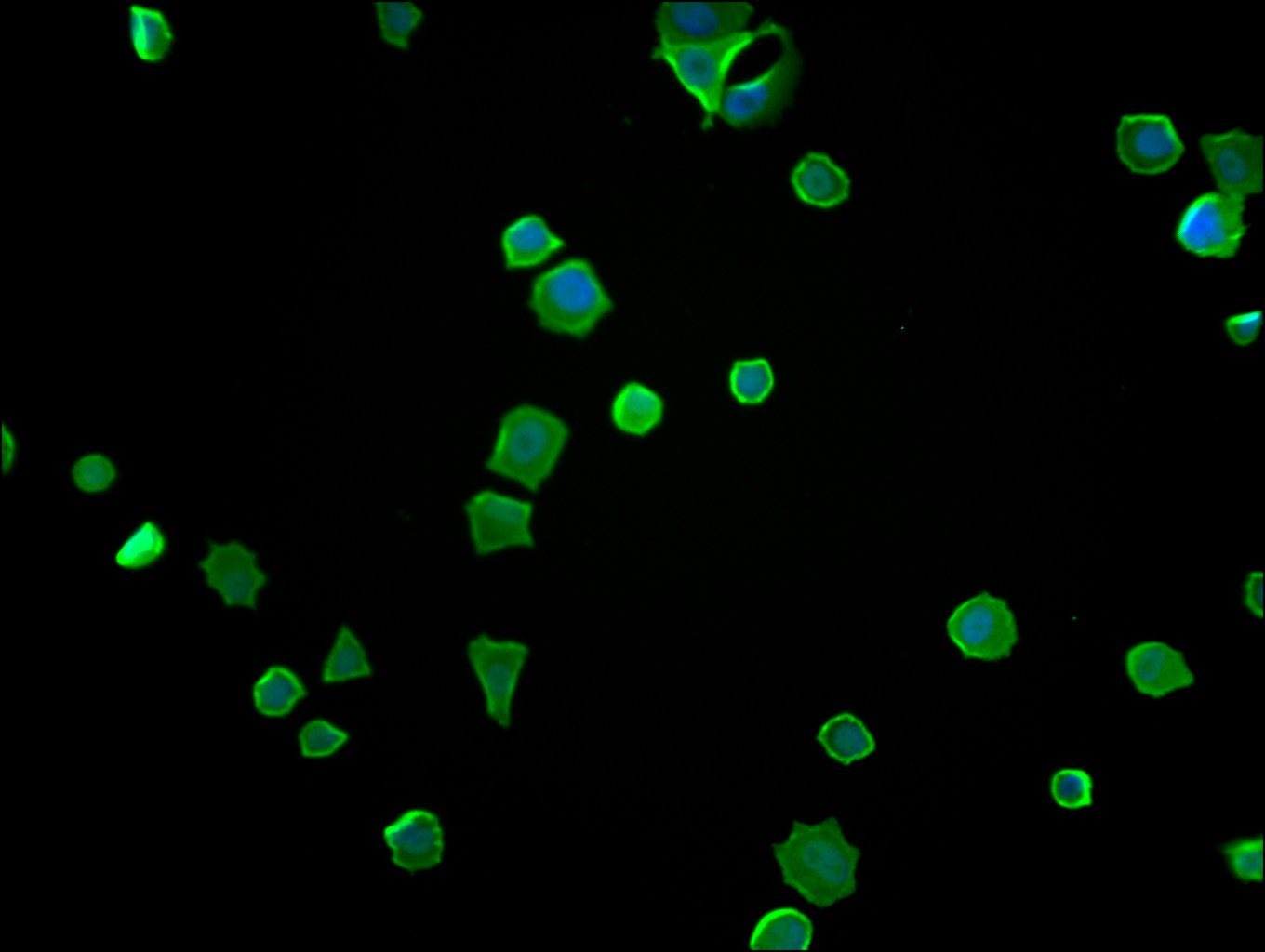
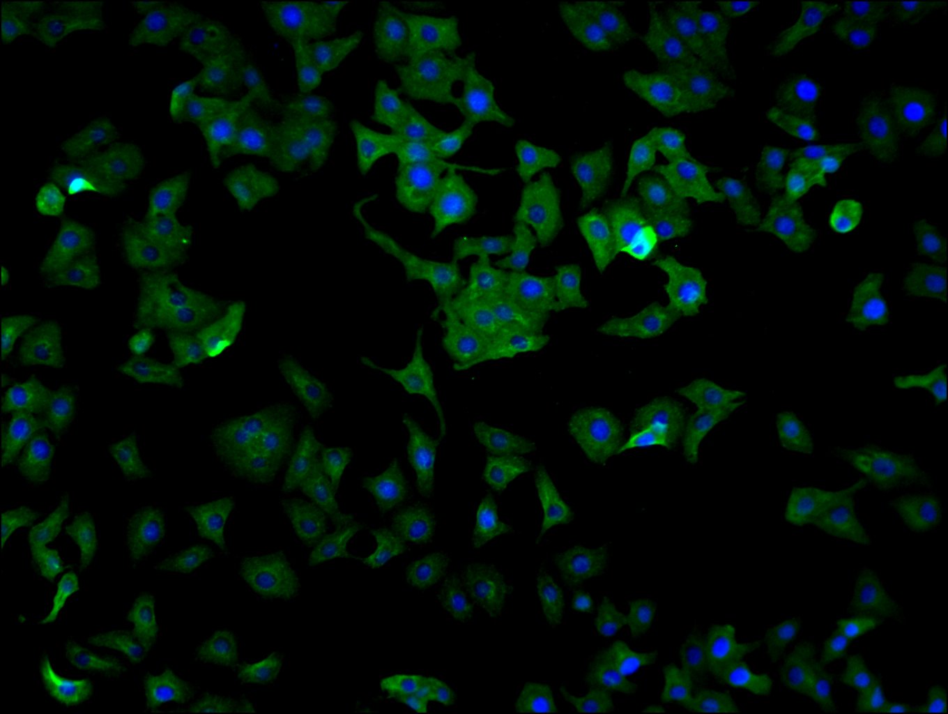
Immunofluorescence staining of MCF-7 cells with CSB-MA007670A0m at 1:130, counter-stained with DAPI. The cells were blocked in 10% normal Goat Serum and then incubated with the primary antibody overnight at 4°C. The secondary antibody was Alexa Fluor 488-congugated AffiniPure Goat Anti-Mouse IgG(H+L).
-
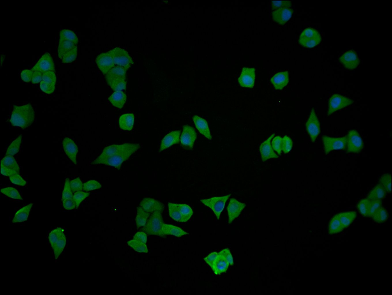
Immunofluorescence staining of Hela cells with CSB-MA007670A0m at 1:130, counter-stained with DAPI. The cells were blocked in 10% normal Goat Serum and then incubated with the primary antibody overnight at 4°C. The secondary antibody was Alexa Fluor 488-congugated AffiniPure Goat Anti-Mouse IgG(H+L).
-
Immunofluorescence staining of HepG2 cells with CSB-MA007670A0m at 1:130, counter-stained with DAPI. The cells were blocked in 10% normal Goat Serum and then incubated with the primary antibody overnight at 4°C. The secondary antibody was Alexa Fluor 488-congugated AffiniPure Goat Anti-Mouse IgG(H+L).
-
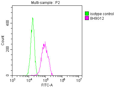
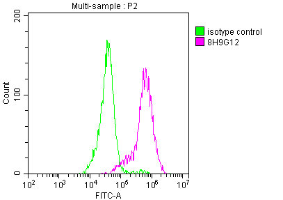
Overlay histogram showing Hela cells stained with CSB-MA007670A0m (red line) at 1:260. The cells were incubated in 1x PBS /10% normal goat serum to block non-specific protein-protein interactions followed by primary antibody for 1 h at 4°C. The secondary antibody used was FITC goat anti-mouse IgG(H+L) at 1/200 dilution for 1 h at 4°C. Isotype control antibody (green line) was used under the same conditions. Acquisition of >10,000 events was performed.
-
Overlay histogram showing MCF-7 cells stained with CSB-MA007670A0m (red line) at 1:260. The cells were incubated in 1x PBS /10% normal goat serum to block non-specific protein-protein interactions followed by primary antibody for 1 h at 4°C. The secondary antibody used was FITC goat anti-mouse IgG(H+L) at 1/200 dilution for 1 h at 4°C. Isotype control antibody (green line) was used under the same conditions. Acquisition of >10,000 events was performed.
-
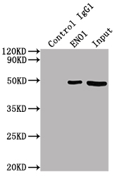
Immunoprecipitating ENO1 in HepG2 whole cell lysate
Lane 1: Mouse control IgG (1µg) instead of CSB-MA007670A0m in HepG2 whole cell lysate. For western blotting, a HRP-conjugated Protein G antibody was used as the secondary antibody (1/2000)
Lane 2: CSB-MA007670A1m (2µl) + HepG2 whole cell lysate (500µg)
Lane 3: HepG2 whole cell lysate (10µg)
-
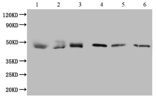
1. Exosomes extracted from plasma
2. Exosomes extracted from serum
3. Exosomes extracted from urine
4. Exosomes extracted from Hela cells
5. Exosomes extracted from latex
6. Exosomes extracted from saliva
-
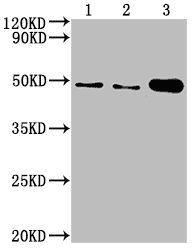
1.Exosomes extracted from MG63 cells
2. Exosomes extracted from Ntera-2 cells
3. MG63 cell Lysate
-
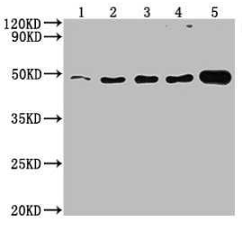
1.Exosomes extracted from HEPG2 cells
2. Exosomes extracted from PC-3 cells
3. Exosomes extracted from Hela cells
4. Exosomes extracted from U87 cells
5. Hela cell Lysate
-
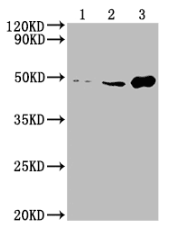
1. Exosomes extracted from Raji cells
2. Exosomes extracted from U251 cells
3. Raji cell Lysate