Call us
301-363-4651 (Available 9 a.m. to 5 p.m. CST from Monday to Friday)
Human neurogenesis takes place mostly during the embryonic, fetal, and neonatal phases, and results in a vast array of neural cell types that make up the human nervous system. The complexity of the brain, a paucity of access to human embryonic and fetal tissues, and the related ethical issues make studying human neurodevelopmental diseases extremely challenging. Experimental models of the human brain are needed for a deeper understanding of its development and diseases. This issue has not made a great breakthrough until Lancaster et al. established the first human brain organoid that partially simulated the 3D structure of the human brain in 2013 [1]. The emergence of brain organoid culture technology provides a very bright way to break through the experimental inaccessibility of the initial stages of the living human brain.
Human brain organoids derived from human pluripotent stem cells (hPSC) recapitulate characteristics of early human nervous development in vitro, including the generation, proliferation, and differentiation of neural progenitors into neurons and glial cells and the complex interactions among the diverse, emergent cell types of the developing brain in three-dimensions [2]. Brain organoids are mainly made up of neural lineage cells, such as neural stem cells, neurons, astrocytes, and oligodendrocytes, and recapitulate human neurogenesis at the cellular level. They provide an experimentally feasible system to visualize and study aspects of human neurodevelopment in a dish. Experts will have a deeper understanding of genetics through brain organoids. With every step forward, breakthroughs are possible to make in more disease areas, transforming physiological and pathological relevance into causation. Human brain organoids can be used to study brain development processes and neurological diseases as well as regeneration.
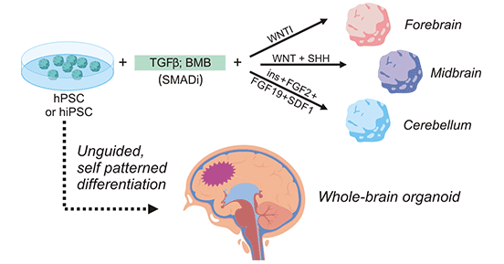
Figure 1. Process of establishment of human brain organoid
https://www.frontiersin.org/articles/10.3389/fcell.2020.590119/full
Scientists have successfully grown organoids similar to the human brain, but previous models have failed to develop human-like functional neural networks. Cleber A. Trujillo et al. designed a better way to culture human stem cells to induce the formation of some neurons in the outer layer of the brain. They found that brain waves generated from brain organoids grown from human stem cells became more complex as development progressed and formed functional neural circuits in miniature brains [3]. And these brain waves share certain features in the developing brains of human babies. The team aimed to further improve organoids and used them to understand disorders associated with neural network failures, such as autism, epilepsy, and schizophrenia
In-Hyun Park and his colleagues found a way to break through the problem of no vascular network existing in human brain organoids. They successfully formed a complex functional network of blood vessels in brain organoids through ectopic expression of the transcriptional factor ETV2 in human cortical organoids (hCOs) [4]. They also showed that vascularized hCOs (VhCOs) differentiated into more complex structures and that neurons are more mature. It was also for the first time discovered the blood-brain barrier (blood-brain barrier, BBB) similar structure to these VhCOs.
Although human brain organoids do partially produce the human brain structure in vitro, each one is unique and they are plagued by high organoid-to-organoid variability [5] [6]. This means they can't be easily used to compare the difference between diseased and normal brain tissue. And this also has raised doubts about whether the developmental process of the human brain can take place outside the context of embryogenesis with a degree of reproducibility comparable to that of endogenous tissue. Silvia Velasco et al. used a specific combination of stem cells to continuously grow multiple human brain organoids in the same developmental order and tissue structure [7]. These cultivated organoids have the same cellular composition and basic connections, and they can survive for a long time in vitro culture environment, and differentiate into a variety of cell types that make up the cerebral cortex, making it possible for researchers to use these organoids for comparative experimental research and drug screening.
SARS-CoV-2 mainly attacks the respiratory tract, but some infected patients have some neurological-related symptoms, including headache, loss of smell, smell disturbance, confusion, epilepsy, and encephalopathy. However, there is no direct experimental evidence that SARS-CoV-2 infects the human central nervous system (CNS). To explore the direct involvement of SARS-CoV-2 in the central nervous system in physiologically relevant models, Bao-Zhong Zhang and his team evaluated SARS-CoV-2 infection in human neural progenitor cells (hNPCs), neurospheres, and induced pluripotent stem cells (iPSCs)-derived brain organoids [8]. The results showed that iPSC-induced hNPCs presented an open state for SARS-CoV-2 infection. Extensive protein expression and infectious virus particles were detected in neurospheres and brain organoids infected with SARS-CoV-2, suggesting that SARS-CoV-2 can efficiently infect the human brain. This study provided the first evidence of direct infection with SARS-CoV-2 in human brain organoids, which could help experts understand the pathogenesis of neurological complications of COVID-19.
Jay Gopalakrishnan's team successfully induced bilaterally symmetrical optic cups in brain organoids by modifying the culture conditions for converting iPSCs into neural tissue and found that this structure can sense light while sending signals to the brain in other areas [9]. When these organoids grow for 50-60 days, the original "eyes" develop into one or two mature visible optic vesicle structures, which are called optic vesicle brain organoids (OVB-organoids). These OVB organoids currently only survive for 60 days. This study first functionally integrates retinal structures into brain organoids, reproducing in an in vitro system the outward extension of nerve fibers from retinal ganglia to connect with target regions of the brain. This system can help to study the "brain-eye" interaction during embryonic development, and provides a powerful tool for the exploration and treatment of retinal diseases, bringing hope for a cure for countless patients suffering from retinal diseases.
A human brain organoid is a primitive form of a brain, but it is not a real brain. It is just a simplified model made for research. So far, no human brain organoids have been found to have consciousness in the lab. Brain organoids cannot restate behavior or indicate which cell types are involved in a particular behavior pattern. Organoids can only compensate for animal models rather than replace them. What organoids can do is allow researchers to do animal experiments more specifically, or get results from animal experiments more quickly and transmit them quickly to patients in a more targeted way.
References:
[1] Lancaster MA, Renner M, Martin CA, et al. Cerebral organoids model human brain development and microcephaly [J]. Nature. 2013;501(7467):373-379.
[2] Qian Yang, Yan Hong, et al. What Makes Organoids Good Models of Human Neurogenesis [J]? Front. Neurosci., 14 April 2022.
[3] Cleber A. Trujillo, Richard Gao, et al. Complex Oscillatory Waves Emerging from Cortical Organoids Model Early Human Brain Network Development [J]. Cell Stem Cell. 2019 Oct 3; 25(4): 558–569.e7.
[4] Cakir, B., Xiang, Y., Tanaka, Y. et al. Engineering of human brain organoids with a functional vascular-like system [J]. Nat Methods 16, 1169–1175 (2019).
[5] Quadrato, G., Brown, J. & Arlotta, P. The promises and challenges of human brain organoids as models of neuropsychiatric disease [J]. Nat. Med. 22, 1220–1228 (2016).
[6] Quadrato, G. et al. Cell diversity and network dynamics in photosensitive human brain organoids. Nature 545, 48–53 (2017).
[7] Velasco, S., Kedaigle, A.J., Simmons, S.K. et al. Individual brain organoids reproducibly form cell diversity of the human cerebral cortex [J]. Nature 570, 523–527 (2019).
[8] Bao-Zhong Zhang, Hin Chu, et al. SARS-CoV-2 infects human neural progenitor cells and brain organoids [J]. Cell Res. 2020 Oct; 30(10): 928–931.
[9] Elke Gabriel, Walid Albanna, Jay Gopalakrishnan, et al. Human brain organoids assemble functionally integrated bilateral optic vesicles [J]. Cell Stem Cell 28 (10): 1740-1757.e8, 2021.
Most Common Organoids
Related Articles